Home
/ Plant Cell Diagram Under Microscope : What Is A Diagram Of A Plant And Animal Cell Under An Electron Microscope Quora - In truth, there are still features of plant and animal cells we're only lately.
Plant Cell Diagram Under Microscope : What Is A Diagram Of A Plant And Animal Cell Under An Electron Microscope Quora - In truth, there are still features of plant and animal cells we're only lately.
Plant Cell Diagram Under Microscope : What Is A Diagram Of A Plant And Animal Cell Under An Electron Microscope Quora - In truth, there are still features of plant and animal cells we're only lately.. Dreamstime is the world`s largest stock photography community. Diagram of plant cell wall. Examining plant cells under the microscope. Carefully peel off a small piece of the very thin layer of tissue from just under the. Robert hooke was the first cytologist to identify cells under his microscope in 1665.
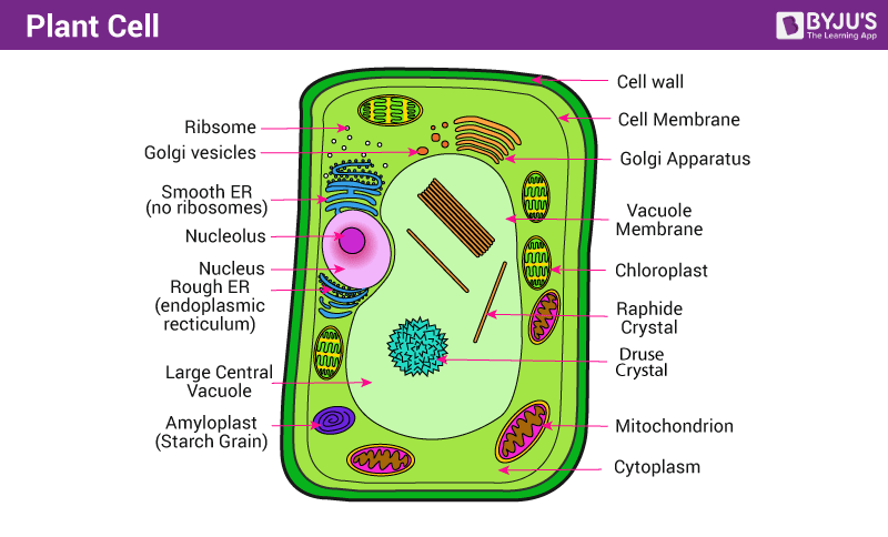
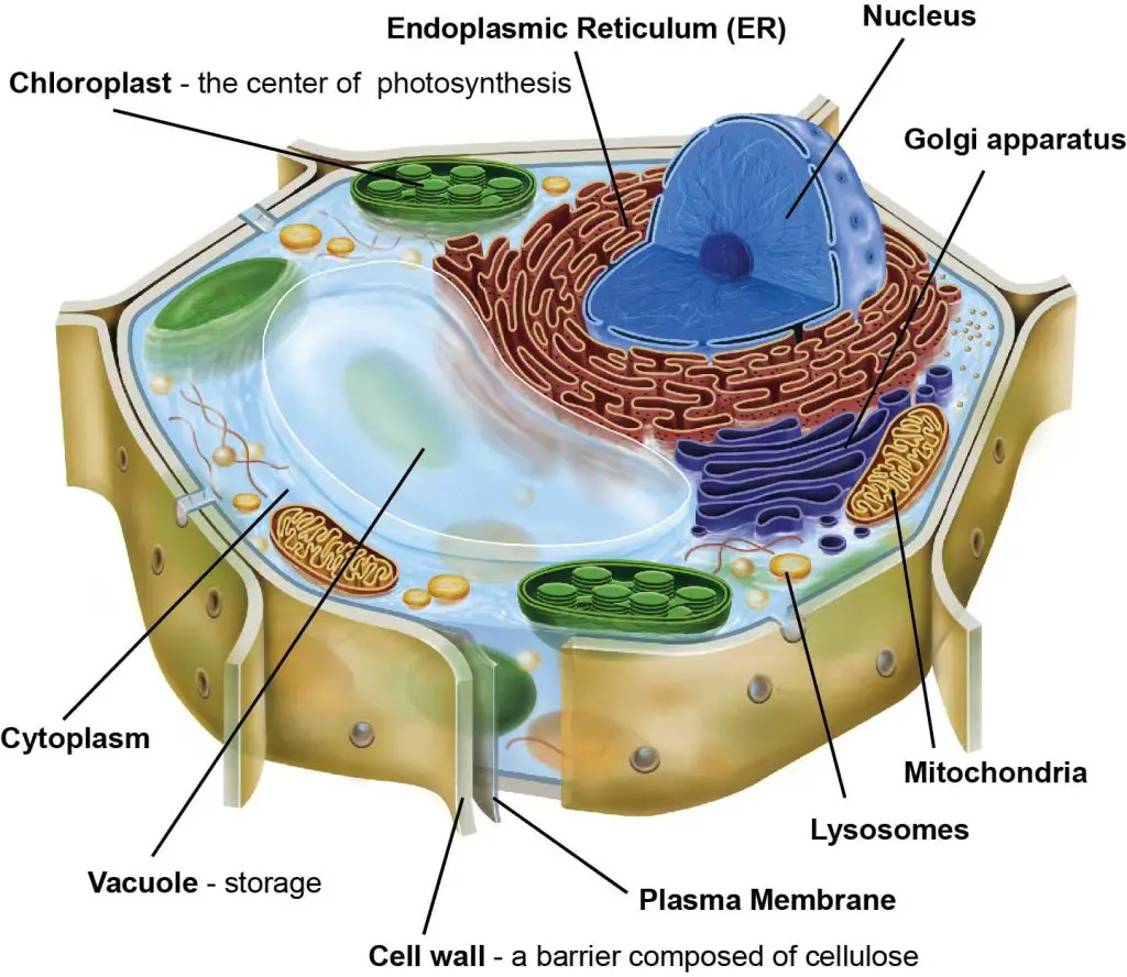
To make observations and draw scale diagrams of cells. Plant cells contain many organelles such as ribosomes, the nucleus, the plasma membrane, the cell wall, mitochondria, and chloroplasts. Robert hooke was the first cytologist to identify cells under his microscope in 1665. Their distinctive features include primary cell walls containing cellulose, hemicelluloses and pectin, the presence of plastids with the capability to perform photosynthesis and store starch. Observe the onion skin under low power of the microscope and then under high power.
Plant Cell Definition Structure Function Diagram Types from cdn1.byjus.com Robert hooke was the first cytologist to identify cells under his microscope in 1665. Plant cells are eukaryotic cells present in green plants, photosynthetic eukaryotes of the kingdom plantae. (ii) presence of large central vacuole in plant cell. They are cells that have a distinct nucleus and other cellular organelles under the microscope, it shows many different parts. The differences between plant and animal cells. A cell is the basic unit of life in all organisms. Examining specimens under a good microscope enables us to study these cellular structures and investigate their biological functions. Epidermal cells include several types of cells that make up the epidermis of plants.
Here's a photo of a plant cell under an electron microscope.
The xylem is responsible for keeping a plant hydrated by transporting water upward from the roots. Your plant cells under microscope stock images are ready. A cell is the basic unit of life in all organisms. Plant cells have cell walls, one large vacuole per cell, and chloroplasts, while animal cells will have a cell membrane only. This lesson is suitable to both junior secondary integrated science (502) and biology (5090) for senior secondary in zambia. The diagram is very clear, and labeled the diagram is very clear, and labeled; Are plant and animal cells the same? But at the same time it is interpretive. They are cells that have a distinct nucleus and other cellular organelles under the microscope, it shows many different parts. Plant cells are the basic unit and building blocks of life in organisms of the kingdom plantae. Observe the labeled diagram of plant. Note that they are composed of phospholipid molecules and protein. Their distinctive features include primary cell walls containing cellulose, hemicelluloses and pectin, the presence of plastids with the capability to perform photosynthesis and store starch.
Ribosomes were first observed under electron microscope by g.e. Share to twitter share to facebook share to pinterest. Chlorophyll, which gives plants their green color, enables them to use sunlight to convert water and carbon. A typical plant cell organelles include cell wall, cell membrane, cytoskeleton, plasmodesmata, chloroplast, vacuoles, endoplasmic reticulum, golgi bodies, mitochondria, ribosomes, peroxisomes, nucleus, nucleolus. Robert hooke was the first cytologist to identify cells under his microscope in 1665.
Lesson 3 Onion Dissection Look At The Plant Cells Rs Science from rsscience.com Light microscopes using visible light and lenses to form a magnified image of the object under investigation e.g. Use them in commercial designs under lifetime, perpetual & worldwide rights. Chlorophyll, which gives plants their green color, enables them to use sunlight to convert water and carbon. Observe the labeled diagram of plant. Although they serve a number of important functions, their primary role is to protect from a variety of harmful factors (environmental stressors) including microbes, chemical compounds as well as ultraviolet light among. Drawing of the structure of cork as it appeared under the microscope to robert hooke from micrographia which is the origin of the word cell. Given below is the diagram of a cell as seen under the microscope after having been placed in a solution A typical plant cell organelles include cell wall, cell membrane, cytoskeleton, plasmodesmata, chloroplast, vacuoles, endoplasmic reticulum, golgi bodies, mitochondria, ribosomes, peroxisomes, nucleus, nucleolus.
Drawing of the structure of cork as it appeared under the microscope to robert hooke from micrographia which is the origin of the word cell.
In addition, plant cells differ from animal cells in a number of key ways. Observe the labeled diagram of plant. Ribosomes were first observed under electron microscope by g.e. The diagram below is a plant cell as may be seen using a light microscope. Plant cells have cell walls, one large vacuole per cell, and chloroplasts, while animal cells will have a cell membrane only. Plant cells are the basic unit and building blocks of life in organisms of the kingdom plantae. A cell is a very tiny structure which exists in living bodies. Cells of plant or animal tissue. Each part, known as an organelle, works together to keep the cell functional. Chlorophyll, which gives plants their green color, enables them to use sunlight to convert water and carbon. Note that they are composed of phospholipid molecules and protein. Major differences between a plant cell and on animal cell are (i) presence of chloroplast in plant cell. Your plant cells under microscope stock images are ready.
Ever since the first microscope was used, biologists have been ch lab # objective: Observe the onion skin under low power of the microscope and then under high power. Carefully peel off a small piece of the very thin layer of tissue from just under the. A typical plant cell organelles include cell wall, cell membrane, cytoskeleton, plasmodesmata, chloroplast, vacuoles, endoplasmic reticulum, golgi bodies, mitochondria, ribosomes, peroxisomes, nucleus, nucleolus. Structure of a plant cell.
A Tour Of The Cell View As Single Page from www.open.edu Each part, known as an organelle, works together to keep the cell functional. Once slides have been prepared, they can be examined under a microscope. Examining a diagram of the plant cell will help make the differences clearer. Plant cells contain many organelles such as ribosomes, the nucleus, the plasma membrane, the cell wall, mitochondria, and chloroplasts. Plant cell is an eukaryotic cell primarily involved in photosynthesis and having its genomic content present in a membrane bound cell organelle, i.e some of these differences can be clearly understood when the cells are examined under an electron microscope. Epidermal cells include several types of cells that make up the epidermis of plants. (ii) presence of large central vacuole in plant cell. (iii) presence of cell wall.
(image uploaded in the folder).
Animal cells also have a because only plant cells perform photosynthesis, chloroplasts are found only in plant cells. The xylem is responsible for keeping a plant hydrated by transporting water upward from the roots. Here's a photo of a plant cell under an electron microscope. Microscopes are needed to study cells in detail. Once slides have been prepared, they can be examined under a microscope. Your plant cells under microscope stock images are ready. Light microscopes using visible light and lenses to form a magnified image of the object under investigation e.g. The diagram is very clear, and labeled the diagram is very clear, and labeled; Eukaryotic plant cell (with diagram). From wikimedia commons, the free media repository. Here's a diagram of a plant cell: Below is a diagram of a part of the plasma membrane. To make observations and draw scale diagrams of cells.
Share :
Post a Comment
for "Plant Cell Diagram Under Microscope : What Is A Diagram Of A Plant And Animal Cell Under An Electron Microscope Quora - In truth, there are still features of plant and animal cells we're only lately."


Post a Comment for "Plant Cell Diagram Under Microscope : What Is A Diagram Of A Plant And Animal Cell Under An Electron Microscope Quora - In truth, there are still features of plant and animal cells we're only lately."