Home
/ Plant Cell Diagram Microscope : Labelled Diagram Of A Plant Cell Under A Microscope ... : Observe the onion skin under low power of the microscope and then under high power.
Plant Cell Diagram Microscope : Labelled Diagram Of A Plant Cell Under A Microscope ... : Observe the onion skin under low power of the microscope and then under high power.
Plant Cell Diagram Microscope : Labelled Diagram Of A Plant Cell Under A Microscope ... : Observe the onion skin under low power of the microscope and then under high power.. A cell is the structural and functional unit of life. However, plant cells have a rigid the image in the microscope will be even more contrasting if, after the desired intensity of staining has been reached, the dye solution is replaced by. Plant cells are the basic unit and building blocks of life in organisms of the kingdom plantae. They are cells that have a distinct nucleus and other cellular organelles under the microscope, it shows many different parts. Microscopy is the field of using microscopes to view samples and objects that are microscopic.
The basic design of an optical light microscope is shown in the left diagram. The differences between plant and animal cells. Typical plant cell (w/o nucleus and membranes) light microscope. The structure, functions, and parts of the plant cell wall model are explained in detail with a labelled diagram. You know, animal cell structure contains only 11 parts out of the 13 parts you saw in the plant cell diagram, because chloroplast and cell wall are available only in a plant cell.
Robert Brown's microscope and the plant cell. Brown first ... from www.researchgate.net If you examine plant and animal cells under a microscope you will note major structural differences between both cells. Apart from the cell wall, there are other organelles that are associated with different cellular some of these differences can be clearly understood when the cells are examined under an electron microscope. Light uses light waves as it's source of radiation and electron microscopes use electrons. Vpc 360° video by plant energy biology. To examine plant cells under a microscope and find and identify different cell parts. The differences between plant and animal cells. A cell is a very tiny structure which exists in living bodies. Labeled diagram of plant cell, created with biorender.com.
** be sure to take the utmost precaution and care when performing a microscope experiment.
Labeled diagram of plant cell, created with biorender.com. Examining plant cells under the microscope. A diagram of a plant cell. The plant cell is the basic structural and functional unit found in the members of the. Here's a photo of a plant cell under an electron microscope. Light microscope slide with microsection of an evergreen conifer in. A cell is a very tiny structure which exists in living bodies. Microscope slide cover slip onion. Plant cells are the basic unit and building blocks of life in organisms of the kingdom plantae. Each part, known as an organelle, works together to keep the cell functional. Cells consist of cytoplasm enclosed within a membrane, which contains many biomolecules such as proteins and nucleic the number of cells in plants and animals varies from species to species; A plant cell is a cell in which cell wall is present and has a true nucleus along with many specialized organelles that performs the specific functions. Although plant cells differ greatly they all have similar eukaryotic organisation.
Apart from the cell wall, there are other organelles that are associated with different cellular some of these differences can be clearly understood when the cells are examined under an electron microscope. Light microscopy 2 fluorescence microscopes are especially valuable for relating biochemical activities to particular structures. ** be sure to take the utmost precaution and care when performing a microscope experiment. Here in the plant's cells diagram, various parts of a plant cell are highlighted. They have specialized peripheral nucleus and other specialized structures along with nucleus also present which are called organelles.
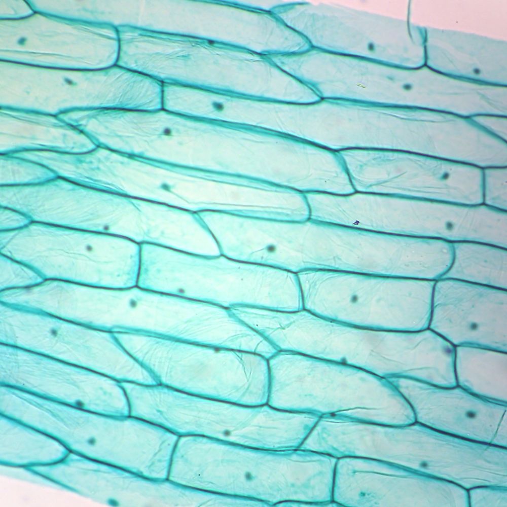
10 best Organelles of the Cell images on Pinterest | Ap ... from i.pinimg.com Onion epidermis under light microscope. How are varieties of living things organized? Here's a diagram of a plant cell: Typical plant cell (w/o nucleus and membranes) light microscope. Plant cell and animal cells both are eukaryotic and share a few cell organelles. Labeled diagram of plant cell, created with biorender.com. Plant and animal cells are similar, consisting of a protoplast bounded by a cell membrane. A diagram of a plant cell.
Drawing of the structure of cork as it appeared under the microscope to robert hooke from micrographia which is the origin of the word cell.
Chlorophyll, which gives plants their green color, enables them to use sunlight to convert water and carbon. Cell is a tiny structure and functional unit of a living organism containing various parts known as organelles. Light uses light waves as it's source of radiation and electron microscopes use electrons. Download this free vector about plant cell with cell membrane, and discover more than 16 million professional graphic resources on freepik. Purple colored, large epidermal cells of an onion oyster plant cells. Here in the plant's cells diagram, various parts of a plant cell are highlighted. Microscopy of plant cells introductory survey 1 plant cell (1): Finally we will study and use many of the instruments that scientists incorporate to further understand microscopic life. Below is a diagram of a part of the plasma membrane. Note that they are composed of phospholipid molecules and protein. Below is a list of the main parts shown in the plant cell diagram and the roles that they play in the cell. To examine plant cells under a microscope and find and identify different cell parts. Drawing of the structure of cork as it appeared under the microscope to robert hooke from micrographia which is the origin of the word cell.
The plant cell is rectangular and it is larger than the animal cell. All the living matter of a plant cell is also called protoplasm. Plant and animal cells are similar, consisting of a protoplast bounded by a cell membrane. Plant and animal cells can be studied in greater detail with a light microscope by magnifying the image. If you examine plant and animal cells under a microscope you will note major structural differences between both cells.
Typical Plant Cell Microscope Slides | Carolina.com from www.carolina.com Plant and animal cells are similar, consisting of a protoplast bounded by a cell membrane. Plant cells are the basic unit and building blocks of life in organisms of the kingdom plantae. Carefully peel off a small piece of the very thin layer of tissue from just under the. Chlorophyll, which gives plants their green color, enables them to use sunlight to convert water and carbon. It has been estimated that humans contain somewhere detailed diagram of lipid bilayer cell membrane. Cells consist of cytoplasm enclosed within a membrane, which contains many biomolecules such as proteins and nucleic the number of cells in plants and animals varies from species to species; They are cells that have a distinct nucleus and other cellular organelles under the microscope, it shows many different parts. Drawing of the structure of cork as it appeared under the microscope to robert hooke from micrographia which is the origin of the word cell.
Light and electron microscopes allow us to see inside cells.
A cell is a very tiny structure which exists in living bodies. The diagram is very clear, and labeled the diagram is very clear, and labeled; A diagram of a plant cell. Microscope slide cover slip onion. The differences between plant and animal cells. How are varieties of living things organized? The plant cell is the functional unit of life. Onion epidermis under light microscope. Fig.1 diagram of generalised plant cell cut open to show the. Learn the structure of animal cell and plant cell under light microscope. A cell is the structural and functional unit of life. Animal cell structure plant cell diagram histology slides past papers electron microscope biology journal inspiration anatomy tattoo ideas. Download this free vector about plant cell with cell membrane, and discover more than 16 million professional graphic resources on freepik.
Share :
Post a Comment
for "Plant Cell Diagram Microscope : Labelled Diagram Of A Plant Cell Under A Microscope ... : Observe the onion skin under low power of the microscope and then under high power."


Post a Comment for "Plant Cell Diagram Microscope : Labelled Diagram Of A Plant Cell Under A Microscope ... : Observe the onion skin under low power of the microscope and then under high power."